What Bones Form The Orbit
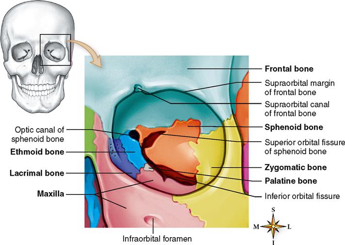
What Bones Form The Orbit - This pyramid, however, is not straight, but displays a laterally tilted axis (black outline in (c) and (d)). The lateral wall comprises the greater wing of the sphenoid bone and zygomatic bone. Sphenoid (cranial) frontal (cranial) ethmoid (cranial) zygomatic (facial) lacrimal (facial) maxilla (facial) palatine (facial) Palatine, zygomatic, lacrimal, and maxilla. Yellow = frontal bone green = lacrimal bone brown = ethmoid bone blue = zygomatic bone purple = maxillary bone aqua = palatine bone red = sphenoid bone teal = nasal bone (illustrated but not part of the orbit) The borders and anatomical relations of the bony orbit are as follows: The cranium is the major portion and it consists of three unpaired bones, the sphenoid, occipital, and ethmoid bones, and three paired bones, the frontal, parietal, and temporal bones. Web the following seven bones form the orbit: Web seven bones form each orbit: Web let's look at how these seven orbital bones join to form different parts of the eye socket (orbit):
Web bones of the orbit and some of the major landmarks. The entrance to the globe anteriorly is approximately 35 mm high and 45 mm wide. Web there are 7 bones that comprise the orbit. Bones, muscles, arteries, veins and nerves this is an anatomy video tutorial covering the. The entrance to the globe anteriorly is approximately 35 mm high and 45 mm wide. Seven bones conjoin to form the. Lesser wing of the sphenoid bone. Web the bones of the orbit develop via both endochondral and intramembranous ossification. Pars orbitalis of the frontal bone lacrimal bone lamina papyracea of the ethmoid bone orbital process of the zygomatic bone orbital surface of the maxillary bone orbital process of the palatine bone greater and lesser wings and body of the sphenoid bone The borders and anatomical relations of the bony orbit are as follows:
Frontal, ethmoid, palatine, lacrimal, maxilla, zygomatic, and sphenoid. Formed by the greater wing of the sphenoid bone and the zygomatic bone. The lateral wall comprises the greater wing of the sphenoid bone and zygomatic bone. The orbital roof is formed by the lesser wing of the sphenoid bone and the frontal bone. Lesser wing of the sphenoid bone. Portions of six bones form its pyramidal walls: The orbital roof is formed by the lesser wing of the sphenoid bone and the frontal bone. Frontal, sphenoid, maxillary, zygomatic, palatine, ethmoid, and lacrimal. Web there are seven bones that contribute to the bony orbit: Web right anterior view of the bony orbit.

The Bony Orbit Borders Contents Fractures TeachMeAnatomy
Frontal, sphenoid, maxillary, zygomatic, palatine, ethmoid, and lacrimal. The sphenoid and ethmoid bones form mostly via endochondral ossification while the frontal bone is formed by intramembranous ossification. Web let's look at how these seven orbital bones join to form different parts of the eye socket (orbit): The lateral wall comprises the greater wing of the sphenoid bone and zygomatic bone..
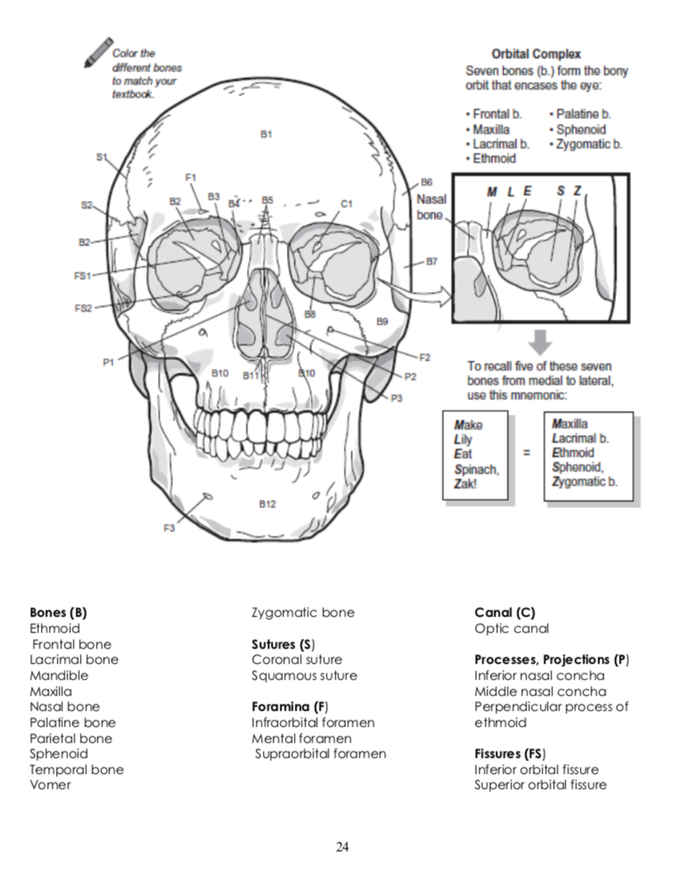
Solved Color the different bones to match your Orbital
Each of these plays a role in keeping the eyeball protected. Lesser wing of the sphenoid bone. However, mri can be a valuable adjunct in certain osseous pathologies especially in determining bone marrow involvement. The lateral wall comprises the greater wing of the sphenoid bone and zygomatic bone. Web the bony orbit and ocular adnexa provide globe protection, allowing normal.

Bones That Form The Orbit / Orbital Bones Ophthalmology Review
The lateral wall comprises the greater wing of the sphenoid bone and zygomatic bone. The orbital roof is formed by the lesser wing of the sphenoid bone and the frontal bone. Web the structure of the orbit is made up of several orbital bones that provide a strong base for the eye so that it can perform its functions properly..

20 best Ophtho images on Pinterest Anatomy, Anatomy reference and
Formed by the greater wing of the sphenoid bone and the zygomatic bone. This pyramid, however, is not straight, but displays a laterally tilted axis (black outline in (c) and (d)). Web seven bones form each orbit: Pars orbitalis of the frontal bone lacrimal bone lamina papyracea of the ethmoid bone orbital process of the zygomatic bone orbital surface of.

Bones of orbit lateral wall Human anatomy and physiology, Human
Zygomatic process of the maxilla and the zygomatic bone zygomatic process of the. Lesser wing of the sphenoid bone. The orbit is a pear shape, with the optic nerve at the stem, and holds approximately 30 cc volume. The orbit is comprised of seven distinct cranial bones. Web key facts about bones of the orbit.

Bones of the orbit Human anatomy and physiology, Anatomy, Orbit anatomy
Frontal, sphenoid, maxillary, zygomatic, palatine, ethmoid, and lacrimal. Although simple, this fact constitutes the basis of the human stereoscopic vision and. Orbital plate of the frontal bone. The frontal, sphenoid, zygomatic, ethmoid, lacrimal, palatine and maxilla bones. The orbital roof is formed by the lesser wing of the sphenoid bone and the frontal bone.
Skeletal System Basicmedical Key
The orbit is comprised of seven distinct cranial bones. The orbital roof is formed by the lesser wing of the sphenoid bone and the frontal bone. The frontal, sphenoid, zygomatic, ethmoid, lacrimal, palatine and maxilla bones. Web the structure of the orbit is made up of several orbital bones that provide a strong base for the eye so that it.

bones that form the orbit Diagram Quizlet
Zygomatic process of the maxilla and the zygomatic bone zygomatic process of the. Web the following seven bones form the orbit: It is our job as ophthalmologists to be able to readily identify these bones and know pretty much every bump, notch, hole, and contour of these bones and what structures pass through, travel along, and attach to these bones..

Orbital Bone Anatomy Human Anatomy Diagram Medical anatomy, Human
Orbital plate of the frontal bone. Maxilla, frontal bone, zygomatic bone, ethmoid bone, lacrimal bone, sphenoid bone, and palatine bone. Sphenoid (cranial) frontal (cranial) ethmoid (cranial) zygomatic (facial) lacrimal (facial) maxilla (facial) palatine (facial) The sphenoid and ethmoid bones form mostly via endochondral ossification while the frontal bone is formed by intramembranous ossification. Optic foramen orbital margin (rim):

Anatomy bones, Orbit anatomy, Anatomy
The orbital roof is formed by the lesser wing of the sphenoid bone and the frontal bone. Each of these plays a role in keeping the eyeball protected. Sphenoid (cranial) frontal (cranial) ethmoid (cranial) zygomatic (facial) lacrimal (facial) maxilla (facial) palatine (facial) Web names of the bones of the orbit with basic anatomy 7 of the cranial and facial bones.
The Depth From Orbital Rim To The Orbital Apex Measures 40 To 45 Mm In Adults.
Bones, muscles, arteries, veins and nerves this is an anatomy video tutorial covering the. Although simple, this fact constitutes the basis of the human stereoscopic vision and. Ct is the modality of choice for orbital bone imaging; Palatine, zygomatic, lacrimal, and maxilla.
Seven Bones Conjoin To Form The.
Lesser wing of the sphenoid bone. It is our job as ophthalmologists to be able to readily identify these bones and know pretty much every bump, notch, hole, and contour of these bones and what structures pass through, travel along, and attach to these bones. Frontal, sphenoid, maxillary, zygomatic, palatine, ethmoid, and lacrimal. There are 7 bones that form the orbit:
The Entrance To The Globe Anteriorly Is Approximately 35 Mm High And 45 Mm Wide.
Web let's look at how these seven orbital bones join to form different parts of the eye socket (orbit): Frontal, ethmoid, palatine, lacrimal, maxilla, zygomatic, and sphenoid. The orbit is comprised of seven distinct cranial bones. Frontal, sphenoid, maxillary, zygomatic, palatine, ethmoid, and lacrimal.
Web Key Facts About Bones Of The Orbit.
Portions of six bones form its pyramidal walls: The orbit is a pear shape, with the optic nerve at the stem, and holds approximately 30 cc volume. The frontal, sphenoid, zygomatic, ethmoid, lacrimal, palatine and maxilla bones. The cranium is the major portion and it consists of three unpaired bones, the sphenoid, occipital, and ethmoid bones, and three paired bones, the frontal, parietal, and temporal bones.